
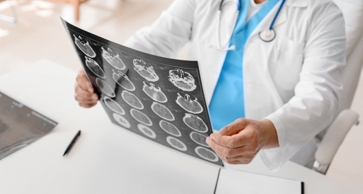
Magnetic Resonance Imaging (MRI) has transformed the world of medical imaging, especially in neurological assessment. This non-invasive technique makes it easier to have images of the brain taken with a higher resolution and is thus used by clinicians for curing different kinds of conditions. In this blog, we shall cover what is MRI on a brain, its advantages, the procedures involved, and the different kinds of conditions that it diagnoses.
What is an MRI?
MRI obtains images of the organs and tissues in the body by applying strong magnetic fields and radio waves. Unlike X-rays or CT scans, MRI does not utilize ionizing radiation; hence, it is safer for most patients. It provides high-resolution images, especially for soft tissue differentiation-a feature well suited for brain imaging.
Why is an MRI of the Brain Performed?
An MRI scan of the brain can lead to the diagnosis of a vast number of diseases and conditions, including the following:
- Tumors: It can observe benign or malignant growth within the brain.
- Stroke: It identifies the sites of ischemia or haemorrhage.
- Multiple Sclerosis: Lesions characteristic of this autoimmune disorder are observed.
- Brain Injuries: The damage caused by trauma on the brain structure is observed.
- Infections: Encephalitis or meningitis, among others, are found.
- Anomalies: Congenital abnormalities or malformations are detected.
- Degenerative Disorders: Assessing a case of Alzheimer’s or Parkinson’s disease.
- The MRI ScanPreparation
Before undergoing the MRI scan, patients are asked to: Take off all metallic objects including necklaces, watches, hair pins, and so on.
Inform them of any implants like pacemakers or cochlear implants which may cause interference with the scans.
Ask for allergies about contrast agents, though most brain MRIs are done without contrast.
During the MRI
- Patient positioning: The patient is then positioned on a moveable table and with his or her head placed in a coil to obtain detailed images of the brain.
- Scanning: You will be slid into the machine by a table, which is a big, round hole that looks like a doughnut. You will lie inside the MRI machine, and you’ll be told not to move when the machine starts scanning. The scans usually last between 20 and 60 minutes.
- Noises: The machine will make noises; these are standard sounds due to the work of the magnets. Should recommend that patients requiring radiotherapy wear earplugs or headphones.
Application of Contrast Agents
Contrast agents of which most are gadolinium-based, are administered through an intravenous for the images to be clearer. It highlights abnormalities that may not be seen in ordinary images.
What Takes Place After the MRI?
Once the imaging is completed, the images are reviewed by the radiologist. After reviewing the images, he or she composes a report and communicates the result to the referring physician. The latter will further communicate the same to his or her patient. Further tests and treatment may be prescribed according to the findings of the MRI.
Advantages of using MRI in Brain Imaging
- Resolution: MRI contrasts well among the areas of brain tissues, and abnormalities may easily be identified more clearly.
- Non-Invasive: MRI is compared to surgical intervention wherein the process is non-invasive, and there is an option to minimize the risks of impacting the patient.
- No Radiation: MRI does not use ionizing radiation. This procedure can be repeated on the patient.
- Functional Imaging: With the help of functional MRI, the assessment of brain function becomes possible by monitoring changes in blood flow.
Risks and Precautions
Although MRIs are in general safe, there are a few concerns:
Claustrophobia: Some patients may develop fear in the confined space of the MRI machine. For such patients, an open MRI machine is available.
Metal Implants: In some cases, implants will make an MRI impossible; hence, a full medical history is important.
Contrast Reactions: Although very rare, some patients develop allergic reactions to contrast agents.
Conclusion
MRI of the brain gives high value since it can accurately diagnose and provide preparation for the treatment of many neurological conditions. Its non-invasive nature, with excellent imaging detail, makes it the first choice of most care providers in health services. It can be helpful and give you an idea of what to expect when you are about to undergo an MRI of the brain: Always refer to your healthcare professional for specific advice and information about your condition.


1 Comment
Your comment is awaiting moderation.
Your point of view caught my eye and was very interesting. Thanks. I have a question for you. https://www.binance.info/register?ref=QCGZMHR6
Your comment is awaiting moderation.
I don’t think the title of your article matches the content lol. Just kidding, mainly because I had some doubts after reading the article.
Your comment is awaiting moderation.
Your article helped me a lot, is there any more related content? Thanks!
Your comment is awaiting moderation.
Thank you for your sharing. I am worried that I lack creative ideas. It is your article that makes me full of hope. Thank you. But, I have a question, can you help me?
Your comment is awaiting moderation.
Your point of view caught my eye and was very interesting. Thanks. I have a question for you.
Your comment is awaiting moderation.
Your article helped me a lot, is there any more related content? Thanks! https://www.binance.info/register?ref=IHJUI7TF
Your comment is awaiting moderation.
Thank you for your sharing. I am worried that I lack creative ideas. It is your article that makes me full of hope. Thank you. But, I have a question, can you help me?
Your comment is awaiting moderation.
Thank you for your sharing. I am worried that I lack creative ideas. It is your article that makes me full of hope. Thank you. But, I have a question, can you help me?
Your comment is awaiting moderation.
Your article helped me a lot, is there any more related content? Thanks!
Your comment is awaiting moderation.
Thank you for your sharing. I am worried that I lack creative ideas. It is your article that makes me full of hope. Thank you. But, I have a question, can you help me?
Your comment is awaiting moderation.
I don’t think the title of your article matches the content lol. Just kidding, mainly because I had some doubts after reading the article.
Your comment is awaiting moderation.
Your point of view caught my eye and was very interesting. Thanks. I have a question for you. https://accounts.binance.com/en/register?ref=JHQQKNKN
I’ve only had contrast once and it felt so bizarre. I would highly recommend talking to your healthcare provider before any similar type of procedure. Just so you know what you’re getting into beforehand.